


- Sku: PT62377
- Vendor: Body of Elements
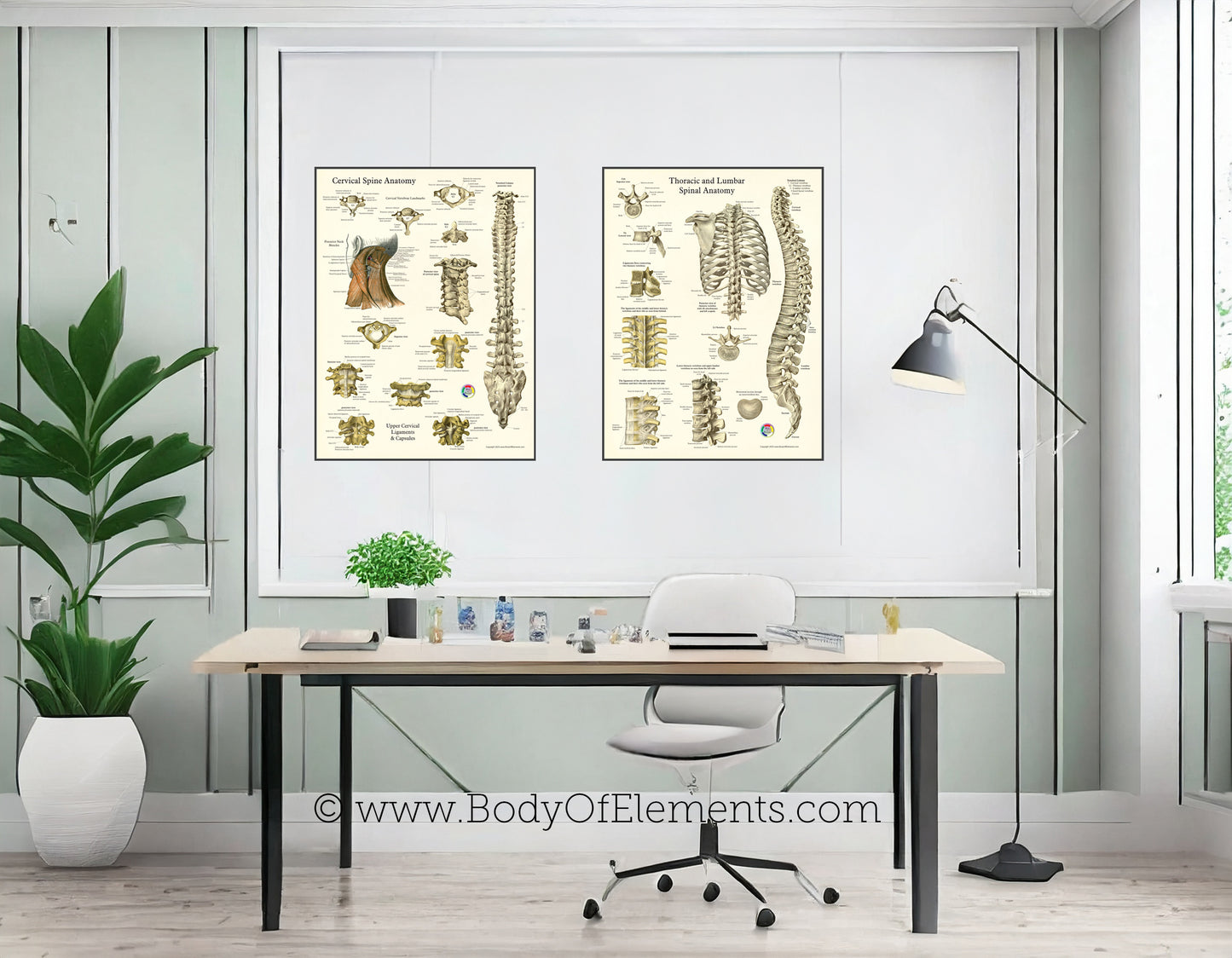
Human Spine and Vertebrae Anatomy Posters
Human Spine and Vertebrae Anatomy Posters 18" X 24"
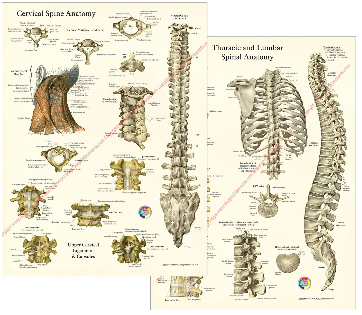
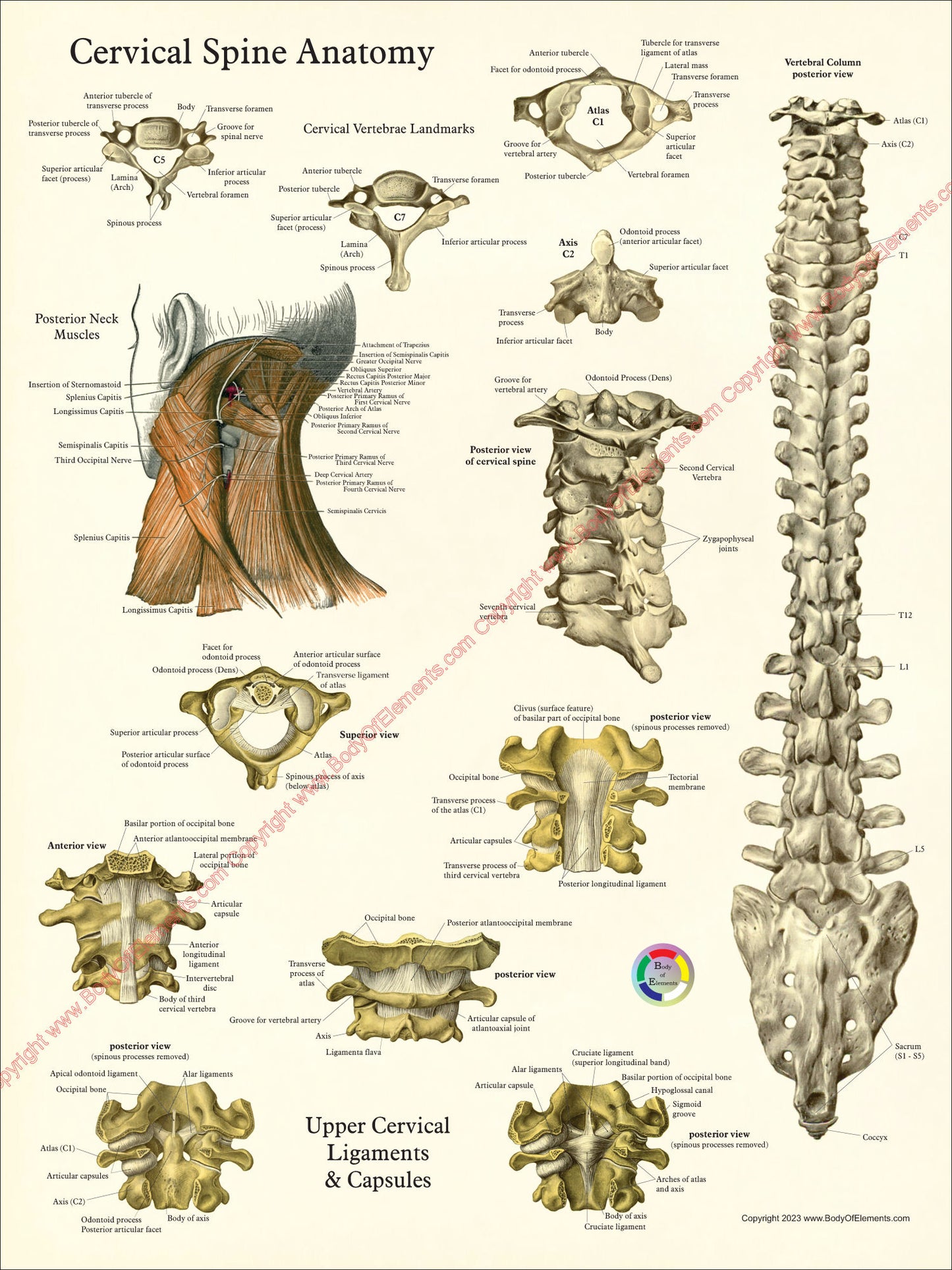
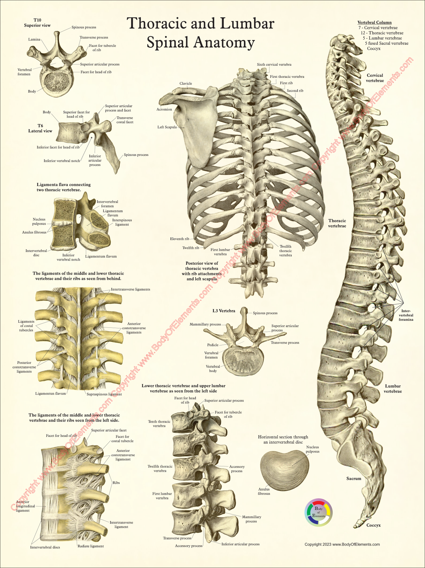
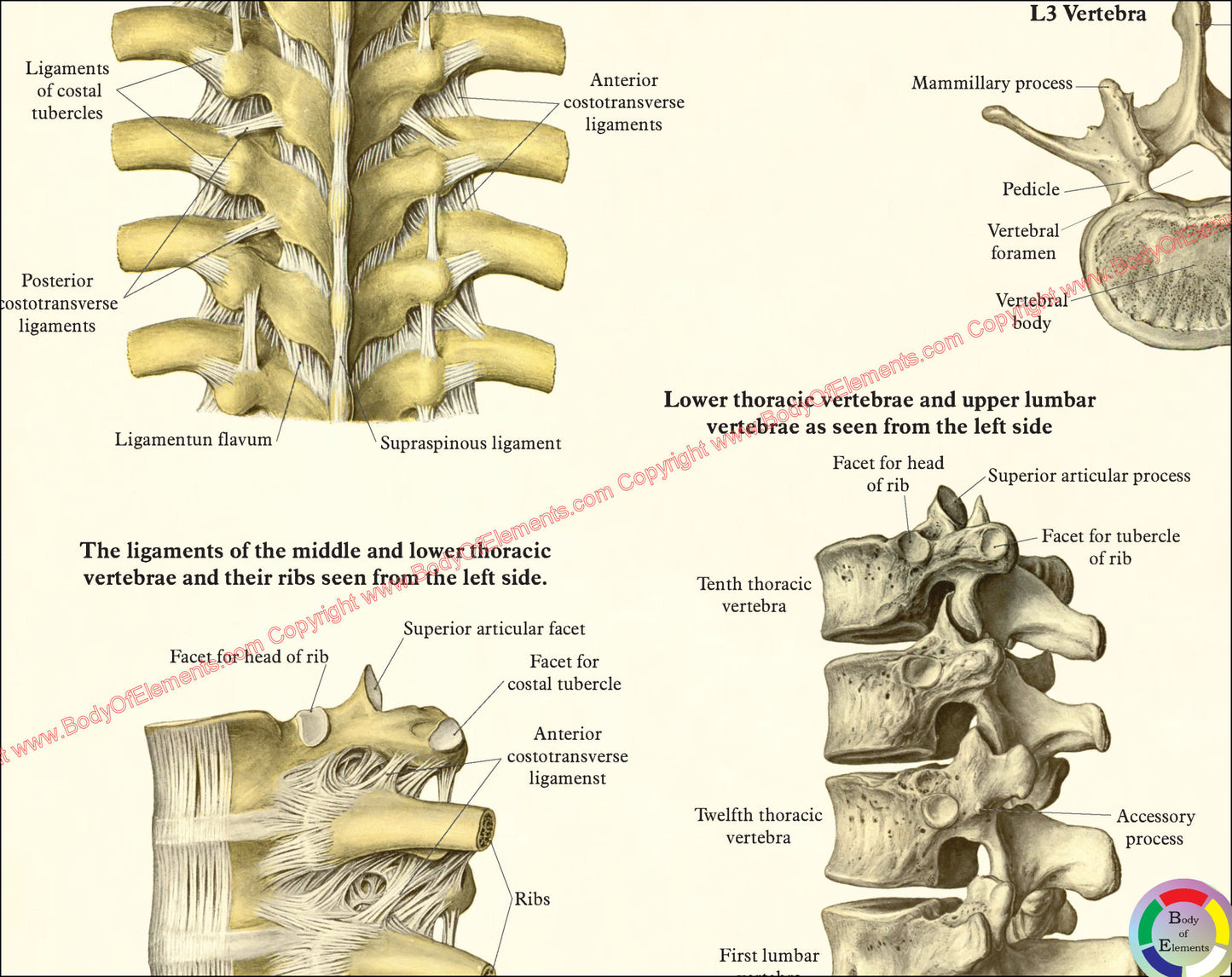
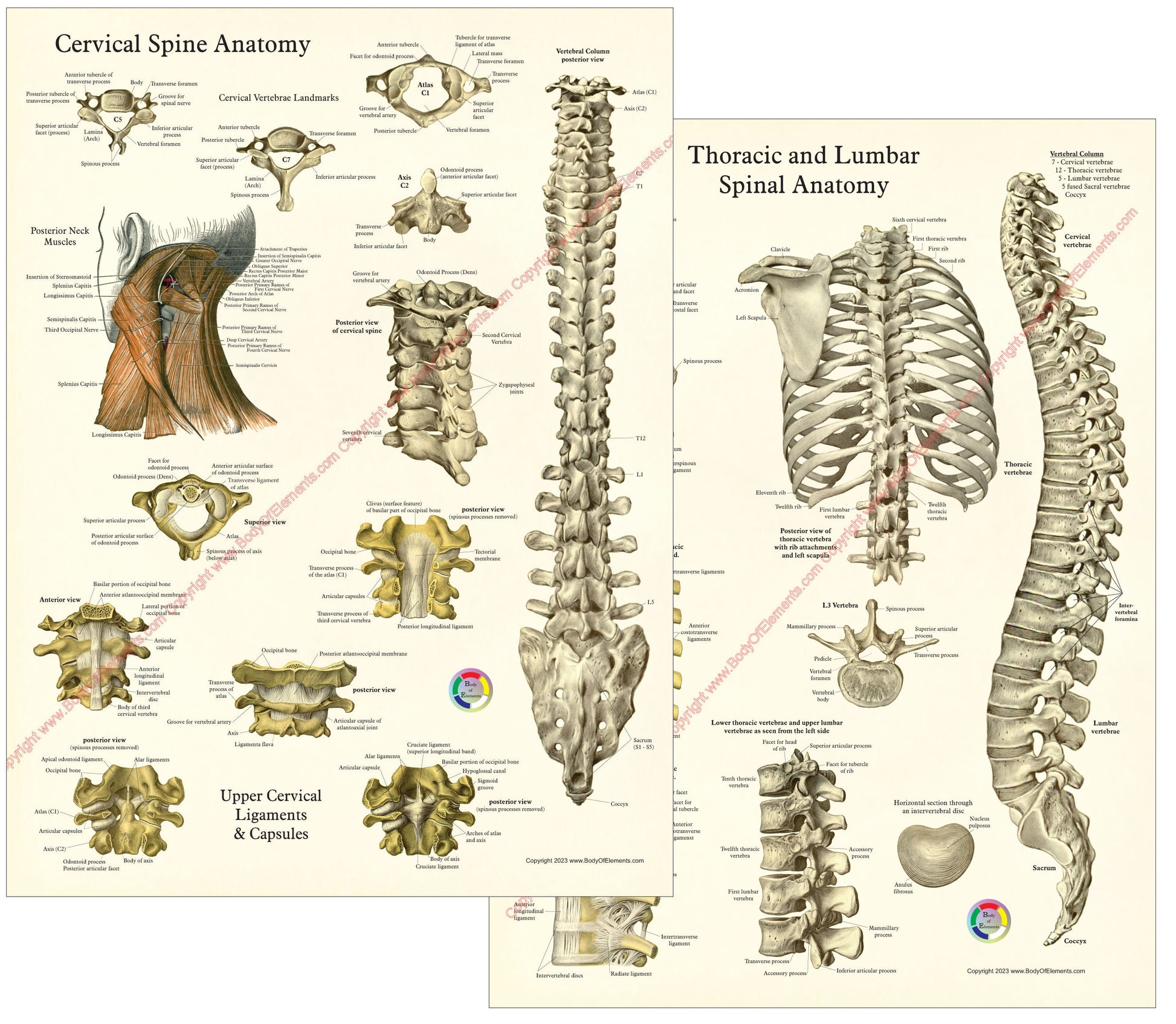
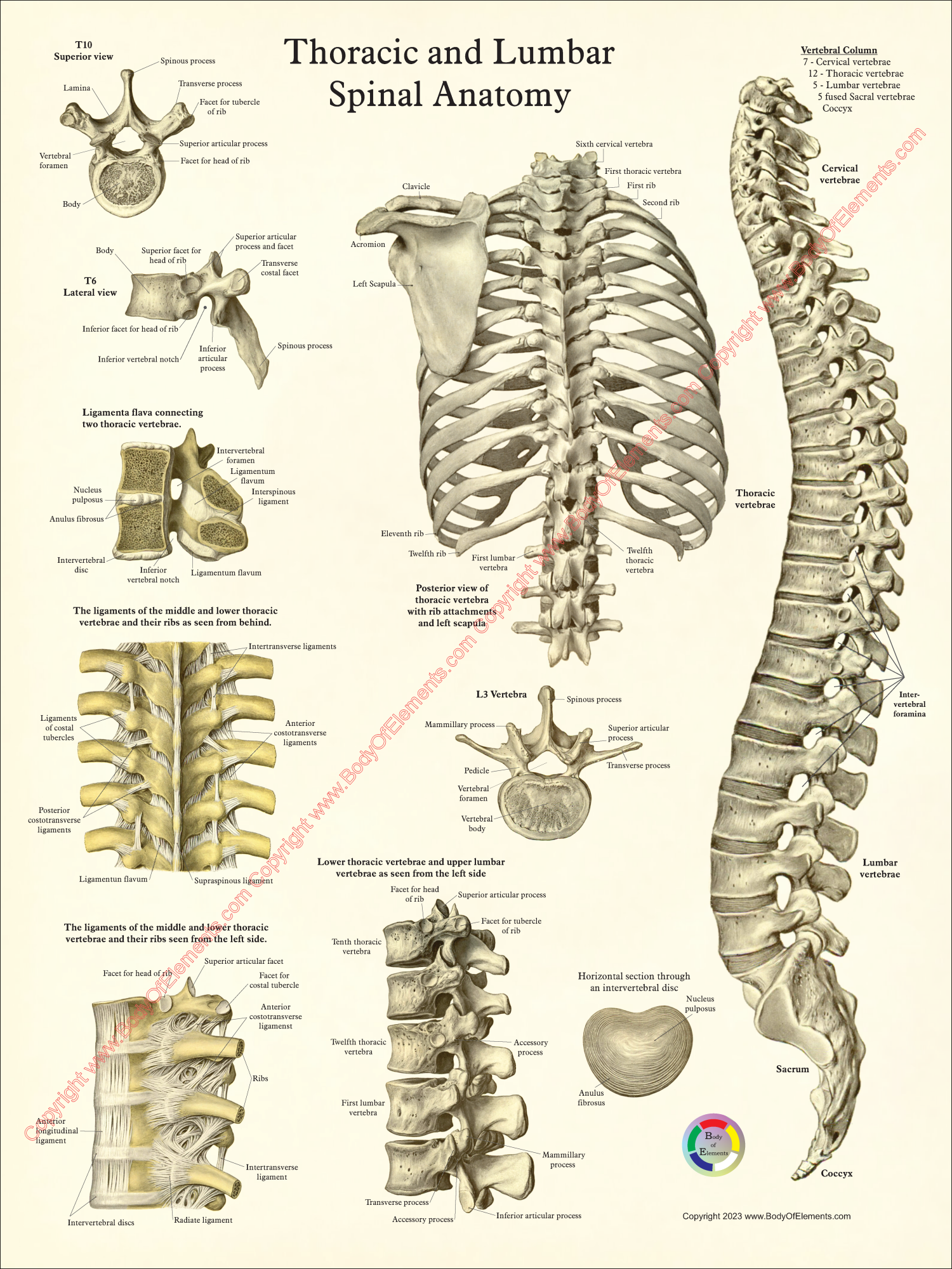
Human spinal anatomy. Two views of the vertebral column: posterior and lateral. Cervical spine with details of landmarks and ligaments of the upper cervicals. Thoracic & Lumbar vertebrae poster showing most common characteristics of the vertebrae along with ligament attachments to the ribs. Nice set of posters to fit any health professionals office, classroom or home for students.
The human spinal column, often referred to as the vertebral column or backbone, is a complex and crucial part of the musculoskeletal system. It serves several essential functions, including protecting the spinal cord, providing structural support, facilitating movement, and serving as an attachment point for various muscles and ligaments. Let's explore the anatomy, joints, and ligaments of the human spinal column:
The human spinal column consists of a series of stacked vertebral bones, separated by intervertebral discs. There are typically 33 vertebrae in total, although they are divided into five regions:
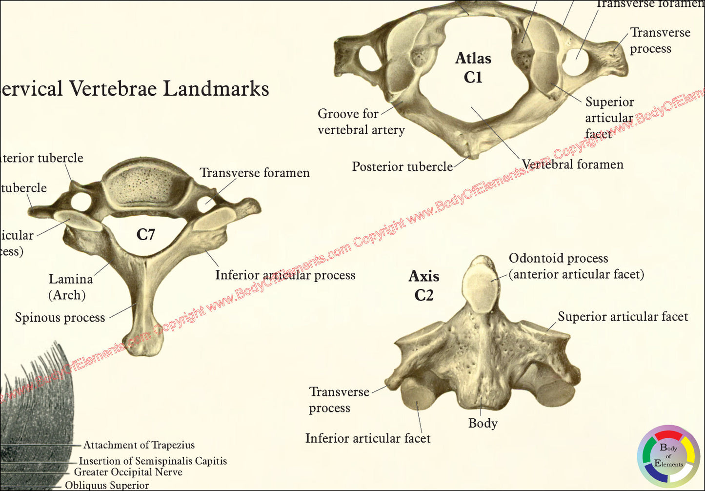
1. Cervical Region: This is the uppermost part of the spine, consisting of seven cervical vertebrae (C1 to C7). The cervical region supports the head and allows for a wide range of neck movements.
2. Thoracic Region: Located in the upper and mid-back, the thoracic region comprises 12 thoracic vertebrae (T1 to T12). These vertebrae connect to the ribs and form the posterior part of the ribcage.
3. Lumbar Region: The lumbar region is in the lower back and is made up of five lumbar vertebrae (L1 to L5). These vertebrae are the largest and support much of the body's weight.
4. Sacral Region: The sacral region consists of five fused sacral vertebrae (S1 to S5) and forms the sacrum, a triangular bone at the base of the spine that connects to the pelvis.
5. Coccygeal Region: The coccygeal region is located at the end of the spine and consists of four fused coccygeal vertebrae. This area is known as the coccyx or tailbone.
Joints of the Human Spinal Column:
The spinal column is made up of several types of joints that allow for different movements and stability:
1. Intervertebral Joints: These are cartilaginous joints found between adjacent vertebral bodies. They allow for limited movement and provide shock absorption. The intervertebral discs act as cushions between these joints.
2. Facet Joints: Also known as zygapophysial joints, facet joints are found at the back of each vertebra. They are synovial joints that facilitate flexion, extension, and rotation of the spine.
3. Atlanto-occipital Joint: This joint connects the first cervical vertebra (atlas) to the base of the skull (occipital bone) and allows for nodding and tilting of the head.
4. Atlantoaxial Joint: Located between the first (atlas) and second (axis) cervical vertebrae, this joint enables the head to pivot from side to side.
This poster is no longer available for purchase directly from our website. You can now order it from our official Zazzle store. - Shop on Zazzle -
Have a question?